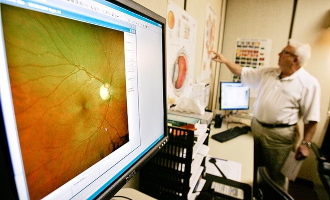
Ever wonder what the inside of your eye looks like?
Well, there’s a whole universe in there, or that’s what it looks like anyway, with a nebulous red and green cloud, and yellow sun.
That yellow sun is your optic nerve and there are blood vessels jagging about around it like a strange electrical storm.
In short: it looks pretty cool.
A device called the Optomap allows you to see a colour snapshot of the inside of your own eye and may one day become common practise during eye exams.
Northern Lights Optometry leased the $275,000 device four years ago and so far has only used it with patients already suffering from various optical ailments.
However, the Optomap retinal exam has proven so useful that Dr. Dick Watts would like to incorporate the device into his routine checkups.
“This is information that we’re going to want to have as part of our routine exams,” said Watts.
“Because it will be standard in practise at some point.”
Watts would like to stress that you don’t necessarily need the retinal exam to detect eye problems.
Using their regular set of tools, including the trusty handheld ophthalmoscope used to peer into the patient’s eye, optometrists are able to detect most eye illnesses during regular checkups.
The retinal exam just makes the physician’s job a little easier.
Using that ophthalmoscope, an optometrist can only see a 15-degree window into your eye at one time.
This is why they’re always asking you to look all around the room: to try to see the eye at different angles.
In some special cases, the patient’s eye can be dilated with drops, giving the doctor a 30- to 40-degree window with which to view the inside of the eye.
But that usually means that the patient has to walk around with dilated pupils for the rest of the day.
The Optomap gets a 200-degree view of the eye, without having to dilate the pupil.
And it takes a picture, allowing the doctor to show the patient exactly what they’re seeing and to keep the picture as a record for next time.
Right now, optometrists take extensive notes and make drawings to remind themselves of what they’re seeing in your eye.
With the Optomap image, the doctor can actually compare the eye over a series of years to track and diagnose potential eye disease.
It also allows northern optometrists to send a full-coloured digital photo to doctors Outside, in cases of referrals.
The Optomap was invented by a Scot named Douglas Anderson, who had a special interest in eye exams.
In 1990, at the age of five, Anderson’s son Leif suffered a spontaneous retinal detachment.
The doctors didn’t detect this detachment until it was too late to treat and Leif lost his sight in one eye.
Anderson blamed the examination techniques, which to him seemed intrusive, crude and - in the case of his son - ineffective.
Anderson put his company, Crombie Anderson Design Consultants, to work researching a new way to view the eye.
He wanted something that was patient-friendly and capable of producing a high-resolution, wide-angled image of the retina.
Four years later, his team had a working prototype and by 1999 it was granted Food and Drug Administration approval in the US.
The Optomap is now used throughout the United Kingdom and North America to help detect problems in the eye including cancers, diabetic retinopathy and retinal holes and detachments.
Using the device is similar to using the other fancy machines that optometrists regularly submit their patients to.
It’s kind of similar to the machine that shoots a puff of air into your eye when you least expect it, only this one doesn’t make you tear up.
You place your eye close to a hole in the machine, aided by the optometrist’s assistant, and stare at a tiny green light.
In a flash of green the picture is taken and appears on a screen next to the machine.
There are actually two flashes, a red and green laser, weak enough to not harm the eye but strong enough to take detailed pictures of different layers of the retina.
It’s painless and, as mentioned above, the images that result can be pretty impressive.
You can spot freckles in the eye (things that optometrists want to watch for growth for fear of melanoma) and even “floaters,” which are harmless inconsistencies in the eye’s vitreous that sometimes float around in your field of vision.
Watts is still unsure when the Optomap will become a permanent part of his routine eye exam, but it could be sometime in late November or early December.
One of the aspects yet to be decided is the price - the fancy retina exams will add to the cost of the checkup.
As of right now, Dr. Watts is trying to convince patients that the extra cost is well worth it.
Contact Chris Oke at
chriso@yukon-news.com