She was an adult black bear — you could tell by her fully-developed and slightly-worn teeth — but she was gaunt, too gaunt, even for May. The gaps between her ribs and the sharp curve of her pelvic bone were visible through her scraggly fur, and her eyes appeared to have sunken deep into her skull.
About nine months after she was put down by conservation officers who found her lurking near a campground, barely responsive to stimuli, the bear’s emaciated body was lying on its side on a metal table in a Whitehorse lab, surrounded by a small group of curious onlookers.
It’s not often the public gets a peek of what goes on at the necropsy lab in the Environment Yukon building at 10 Burns Rd., but on the evening of Jan. 24, about a dozen people got exactly that — and more.
Led by Yukon chief veterinary officer Mary Vanderkop and environment laboratory coordinator Meghan Larivee, the two-hour-long event, organized by Yukon Wildlife Viewing, gave attendees the chance to get up close and personal with the insides and outsides of several Yukon specimens.
“It’s an important part of the work we do and so I think it’s important for us to share that aspect with people,” Vanderkop said.
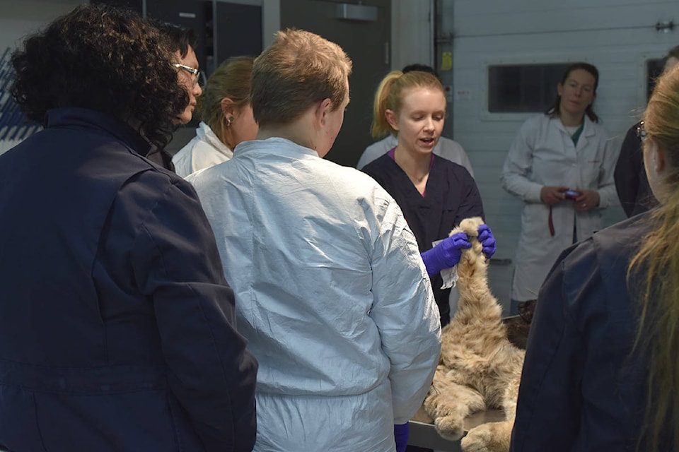
After donning nitrile gloves, lab coats and plastic boot covers, attendees entered the lab, where a strip of yellow tape on the floor divided the “wet” and “dry” sides of the room.
On one counter sat three plastic bins, similar to the ones used at airport security lines, but holding precious cargo that would likely never be accepted as carry-on items: a whole red fox, stiff and slightly curled up; the head of a mule deer, its large eyes glazed over; and what almost looked like a large flesh-coloured pillow with odd stubs sticking out of it that turned out to be a skinned beaver, feet and tail removed.
A raven, wings tucked in, also sat next to the bins, but the main stars of the night were on a metal table about a metre away — the bear, on its side but with a clean cut running down its underside visible, and, in another plastic bin, a young lynx, its fur in pristine condition. Almost all bears killed in the Yukon by human conflict will eventually make their way on to this table, too.
The lynx, Larivee explained, was shot by a farmer who discovered it going after his chickens. She encouraged attendees to run their fingers through the animal’s thick fur and to examine the huge paws — natural snowshoes, an invaluable asset during Yukon winters — but the lynx would not be opened up tonight. The lab is required to skin carcasses if the pelts are in good condition, Larivee said, so they can be used for educational purposes or given to First Nations.
A lab assistant returned the lynx to its spot in the fridge as attention turned to the bear, chunks of its fur missing or matted. Necropsies begin with external examinations, Vanderkop said, guiding attendees’ hands to where the bear’s bones could be easily felt through the skin, and most people prefer having the animal’s head pointing to the left to make visualizing its insides easier.
“It’s a dead body, but it’s treated with respect,” Vanderkop said as she lifted two of the bear’s legs, putting the animal on its back and revealing its rib cage. She pointed out the lack of fat under the skin, a sign the bear was severely malnourished, and also the hole in the skin and rib cage caused by the bullet that killed it.
Vanderkop then used a pair of pruning shears to remove the top of the bear’s ribcage, the crunch of the garden tool cutting through bone and tissue oddly similar to that of cutting through cardboard with scissors. One attendee turned away and left the lab shortly after. Another followed suit after the bear’s glistening organs, in shades of deep red, beige and, in the case of the intestines, a blueish-green, were revealed.
A bear’s internal anatomy closely mirrors that of a human’s, so much so that bears can be used during training exercises for coroners when human cadavers aren’t available. Using a knife, Vanderkop pointed out and began removing key organ structures, starting with the tongue, esophagus, trachea, lungs and heart. Cartilage rings running down the trachea keep the structure from collapsing, Larivee said, ensuring the airway stays open even when put under pressure.
Vanderkop ran a pair of scissors down the trachea. There were no obstructions, she noted, but pointed out some clotted blood, likely inhaled by the bear in its last moments. One of the lungs, squishy to the touch, appeared to have taken the brunt of the shot, fragments of rib visible in the delicate dark-pink structure.
Examining the heart and kidneys, each about the size of a child’s fist, Vanderkop noted that there was were no fat deposits on or around any of those organs, another indication that the bear had been food-deprived for a long time. Bear kidneys are unique in that they have “compartments” within them. When cut in half and then laid flat, little square chunks, almost like a mango half that’s been scored and then turned inside-out, bulge out from the surface.
A quick, clean cut to the bear’s stomach, small and yellowish-beige, revealed her last meagre meals — on one side, a bright red mush, some berries still undigested, and on the other side, a green, fibrous pulp, possibly the remnants of grasses or dandelions. A few green strands were also caught between the bear’s teeth.
The raven necropsied next proved much more well-fed. Fat bulged from the incision that Larivee made just below its sternum, a dark red liver, almost purple, hiding underneath. The bird, Larivee said, was found about a week ago near a hydro pole. Burn marks on the underside of its scaly feet seem to confirm electrocution.
Opening up the raven further, Larivee pointed out its unique lung structure — birds don’t have diaphragms, she explained, and the lungs actually sit in little compartments between their ribs. Birds also have special air sacs connected to their lungs which means they can basically be holding two breaths at once, a useful ability when flying. Vanderkop pointed out a tiny structure located in the birds lower half about the size and shape of a grain of rice — a testicle, she said, adding that birds can’t be sexed from the outside.
In the interview afterwards, Vanderkop said that the information collected from the bear and raven would be entered into reports, and those, in turn, entered into a database, as is done after every necropsy at the lab.
The eagerness of the attendees at the event was encouraging, she said.
“Certainly, the degree of interest and the variety of questions is nice to hear, and I think people are interested, and we’re very interested to share what we do, especially the fact that it is getting the most from the animal, and it is respectful of the wildlife,” she said. “It is not cutting it up for the fun of it.”
Contact Jackie Hong at jackie.hong@yukon-news.com
This story has been updated to correct the spelling of Meghan Larivee’s last name.